Tract-Tracing and Brain Connectivity
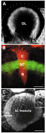
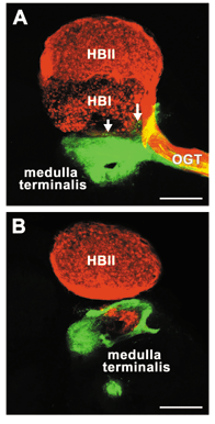
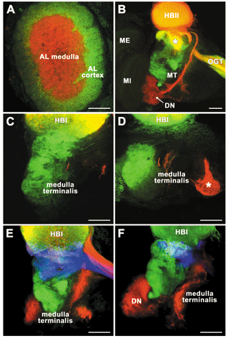
Jeremy Sullivan has conducted a series of gorgeous studies using a injections of lipophilic tracers (DiI and DiA) or dextrans coupled to fluorophores, and immunocytochemical assays in the brains of several crustacean species. The primary goal of this work was to clarify the connectivity among neuropil regions in the deutocerebrum and protocerebrum in crayfish and lobsters, in order to define the pathways that handle olfactory information after primary processing in the olfactory lobes. DiI and DiA injections into the olfactory and accessory lobes in Homarus americanus, Procambarus clarkii and Orconectes rusticus revealed that the cluster 10 projection neurons innervating the accessory lobe exclusively innervate the neuropils of the hemiellipsoid body, while projection neurons innervating the olfactory lobe primarily target the medulla terminalis. These studies demonstrated that the projection neuron pathways from the olfactory and accessory lobes project to separate, largely non-overlapping regions of the lateral protocerebrum (Sullivan and Beltz, 2001a). Additional studies in the lobster showed that the ascending pathways from the antenna II and lateral antenna I neuropils, which are involved in the processing of chemosensory and tactile inputs, innervate the same regions of the medulla terminalis and that these regions are different from those innervated by the olfactory lobe output pathway (Sullivan and Beltz, 200b). Most recently, Jeremy has demonstrated an anatomical segregation of olfactory and multimodal inputs within the accessory lobe and hemiellipsoid body, demonstrating important functional subdivisions within both of these regions (Sullivan and Beltz, 2005). Jeremy Sullivan's work has also contributed to our understanding of the evolution of the olfactory pathway. Using the same type of focal dye injections that were so successful in crayfish and lobsters (Sullivan and Beltz 2001a,b), pathways of four eumalacostracan taxa (Stomatopoda, Dendrobranchiata, Caridea, and Stenopodidea) that diverged from the eureptantian line prior to the appearance of the accessory lobes were examined. The innervation patterns of the olfactory projection neurons of the species examined were found to differ markedly, varying from that observed in the most basal taxon examined (Stomatopoda), in which the neurons primarily project to the medulla terminalis, to those observed in the two highest taxa examined (Caridea and Stenopodidea), in which they primarily target the hemiellipsoid body. These results suggest that substantial changes in the relative importance of the medulla terminalis and the hemiellipsoid body within the olfactory pathway have occurred during the evolution of the Eumalacostraca (Sullivan and Beltz, 2004).
|
Home | Links | Contact Us
- Maintained by: Barbara Beltz & Jeannie Benton
- Created: June 2, 2006
- Last Modified: June 2, 2006
- Expires: June 2, 2006


