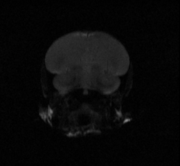
functional Magnetic Resonance Imaging
Functional magnetic resonance imaging (fMRI) is a noninvasive technique that allows the collection of real-time, whole brain data, and is useful in longitudinal studies. fMRI allows us to indirectly observe neural activity during a time course using blood oxygenation as an indirect measure of neuronal activation; fMR images are acquired quickly, images reflect the localized hemoglobin deoxygenation that occurs when neurons respond to a stimulus (Logothetis, 2003, Logothetis and Pfeuffer, 2004) . While fMRI is now commonly used in humans, only relatively recently was this method adapted and optimized for small brains, such as that of the zebra finch, with relatively high regional spatial and temporal resolution to address biological questions (Van Ruijssevelt et al., 2013). Unlike other techniques, such as electrophysiology and immunocytochemistry, fMRI possesses the unique benefit of being able to noninvasively observe a whole brain response to a stimulus in real time.
We are able to introduce multiple auditory stimuli and observe subsequent responses over time in a single subject. Previous fMRI research in zebra finches has cross sectionally corroborated and furthered our understanding of tutor and conspecific song processing in the zebra finch brain and demonstrated how tutor-song specific responses change during normal development (Poirier et al., 2009, Van der Kant et al., 2013, Van der Kant, 2015). Currently, we are using fMRI to study the auditory learning process longitudinally throughout development, specifically observing the changes in neural response before and after a short tutoring experience.
References:
- Van der Kant, A.M. (2015). Neural Correlates of Vocal Learning in Songbirds and Humans: Cross-species fMRI Studies into Individual Differences. Doctoral. Leiden University Center for Linguistics and University of Antwerp.
- Van der Kant, A., Derégnaucourt, S., Gahr, M., Van der Linden, A., and Poirier, C. (2013). Representation of Early Sensory Experience in the Adult Auditory Midbrain: Implications for Vocal Learning. PLoS ONE 8, e61764.
- Logothetis, N.K. (2003). The Underpinnings of the BOLD Functional Magnetic Resonance Imaging Signal. J. Neurosci. 23, 3963–3971.
- Logothetis, N.K., and Pfeuffer, J. (2004). On the nature of the BOLD fMRI contrast mechanism. Magnetic Resonance Imaging 22, 1517–1531.