|
Form – Line
The overall feat of vision is the effective detection of biologically useful information in as efficient a fashion as possible. From an infinite amount of potential visual stimuli, the visual system must select only those elements that are relevant to behavior. For example, the visual system is more sensitive to information that can tell you where an object begins and ends, indicated by discontinuities in the pattern of light falling on the retina, than information that can tell you about the spectral composition or luminance of the light source itself. In fact the perception of such discontinuities—edges—does not change under dim, bright, or even colored light. For instance, the text in a book appears the same under low lamp-light and bright sunlight. Many visual modalities, including color, depth, motion, and luminance show much greater sensitivity to local changes across the visual field than gradual ones—it is these regions of change that carry the most useful information because they demarcate object boundaries, which are most deserving of our visual attention. This sensitivity is achieved by center/surround organization of the receptive fields of the visual system.
Center-surround cells
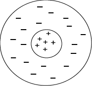
 In 1953 Stephen Kuffler made a critical breakthrough in vision research when he discovered that retinal ganglion cells of the cat could be activated by small spots of light much better than large spots—that is, these cells did not respond well to diffuse light. Further, each ganglion cell responded best to a small spot of light at a particular site on the retina, the cell’s receptive field. The receptive field is determined by the group of photoreceptors that are wired up to the cell in question; these photoreceptors correspond to a restricted region of the visual field. For a given cell, different regions of the receptive field can respond differently to light. Light falling on the center of a retinal ganglion cell’s receptive field may excite the cell while light falling on its surround may inhibit it, through a process termed lateral inhibition—this explains why diffuse light does not activate these cells very well. What a retinal ganglion cell is measuring is the pattern of brightness (relative luminance) across its receptive-field rather than the absolute value of light falling on it (luminance). In 1953 Stephen Kuffler made a critical breakthrough in vision research when he discovered that retinal ganglion cells of the cat could be activated by small spots of light much better than large spots—that is, these cells did not respond well to diffuse light. Further, each ganglion cell responded best to a small spot of light at a particular site on the retina, the cell’s receptive field. The receptive field is determined by the group of photoreceptors that are wired up to the cell in question; these photoreceptors correspond to a restricted region of the visual field. For a given cell, different regions of the receptive field can respond differently to light. Light falling on the center of a retinal ganglion cell’s receptive field may excite the cell while light falling on its surround may inhibit it, through a process termed lateral inhibition—this explains why diffuse light does not activate these cells very well. What a retinal ganglion cell is measuring is the pattern of brightness (relative luminance) across its receptive-field rather than the absolute value of light falling on it (luminance).
Have you figured out why you see illusory grey dots at the intersections of the Hermann grid on the main page yet? The illusion arises due to center/surround cell behavior, but we’ll leave it up to you to put two and two together…
Both retinal ganglion cells and thalamic cells to which they project have center/surround organization.
Edge detection: Oriented cells and extended contour cells
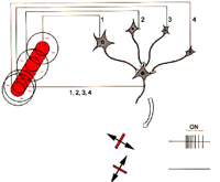
A few years after Kuffler discovered center/surround organization, his students David Hubel and Torsten Wiesel set to work on studying the responses of cells in the primary visual cortex, the next stage of visual processing along the visual pathway. Hubel and Wiesel projected small spots onto a screen using slides with bits of black paper glued to them and recorded responses from cells from the visual cortex of a cat whose eyes were directed at the screen. For a long time Hubel and Wiesel could not find any cells that responded to these spots of light. When they finally did find a somewhat responsive cell, they spent nine hours trying to get a good response out of it. In the final hour, they realized it was not the dark spot on the slide that the cell was responding to, but rather the line of shadow cast by the slide as they moved it in and out of the projector! Moreover, they discovered that the cell responded only when the shadow was oriented horizontally.  Model of the receptive-field of an orientation-selective “simple cell” in primary visual cortex. Adapted from Hubel and Wiesel, 1962.
This pair of Nobel-winning scientists went on to find that V1 contained cells selectively responsive to all possible orientations. Here was the key to edge-detection. Cells in the visual cortex were integrating the responses of groups of thalamic center/surround ganglion cells to extrapolate contours, like a game of connect the dots.
The orientation selectivity of an oriented cortical cell depends on the orientation of the receptive fields of the thalamic cells that make up its receptive field.
To get an extended contour, like a corner, groups of oriented cells must be sampled by cortical cells at the next stage in processing. With every successive stage in visual processing the responses of cells from the previous stage are selectively integrated to extrapolate yet another level of visual complexity over increasingly extensive areas of the visual field.
Line Drawings
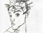
 A line is a discontinuity in color or lightness connecting two points in a two dimensional plane. It is exactly what our center/surround cells are interested in, making it a powerful visual tool when wielded skillfully. Line drawings, even very simple ones, can effectively convey three-dimensionality despite being no more than an assemblage of marks on a two dimensional surface. Effective line drawings are effective because they activate the visual system in a similar way to the real world. By making line drawings, artists since the beginning of art history with cave drawings have recognized something profound about how the structure of visual input relates to subjective experience. Artists cultivate those insights by constantly conducting psychophysical experiments—by making art. A line is a discontinuity in color or lightness connecting two points in a two dimensional plane. It is exactly what our center/surround cells are interested in, making it a powerful visual tool when wielded skillfully. Line drawings, even very simple ones, can effectively convey three-dimensionality despite being no more than an assemblage of marks on a two dimensional surface. Effective line drawings are effective because they activate the visual system in a similar way to the real world. By making line drawings, artists since the beginning of art history with cave drawings have recognized something profound about how the structure of visual input relates to subjective experience. Artists cultivate those insights by constantly conducting psychophysical experiments—by making art.
 Self portrait, Egon Schiele, 19??
 |
If a drawing is said to have perspective, this means it is drawn from the view point of an individual with a visual system, so it should use cues that resonate with subjective experience. It turns out, certain visual phenomena must be conserved to effectively achieve this, while others, like the direction of a shadow on the ground, can be inaccurate and will go unnoticed. For instance, objects that are the same size in absolute terms, but occur at differing distances in the visual field should be represented as different sizes, with the farthest objects vanishing altogether at the horizon—because that’s how we actually see and make judgments regarding the distance of objects. And the lip of a circular vessel should be represented as elliptical, despite being a circular form, because you can’t actually see the circular nature of the opening unless looking at it from above. |
In the course we will examine various consequences of neural processing on art practice.
|