Form – Resolving the Image — Visual Acuity
Depth of Focus: the Lens and the Iris
 |
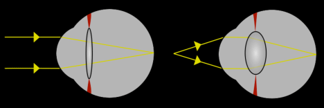
The Human eye contains a transparent biconvex shaped crystalline lens that helps to refract light to be focused on the retina. By changing shape, as cilliary muscles contract and relax, the lens is able to alter the eye's focal length—its ability to converge (focus) or diverge (diffuse) light—so that the eye can focus on objects at differing distances. This is called accommodation. As you get older, your ability to accommodate degrades, which is why older people need reading glasses. |
This figure shows light from a distant object and a near object being focused on the retina by the process of accommodation:
The
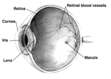
iris is a muscular pigmented structure that forms the eye's
pupil, an opening through which light enters the eye. The iris is responsible for contracting and expanding to regulate the amount of incoming light. It is like the aperture in a camera. Under bright light conditions it contracts, reducing the amount of light let through, and increasing the
depth of focus. When a cone of light enters the eye it is focused such that the individual rays of light converge at the same point. A small point of convergence yields a sharp focused image. A smaller hole yields a smaller point of convergence and in turn a more focused image. However, if the hole is
too small the rays of light converge
before reaching the retina and then begin to spread outward again producing a less focused image. The retinal ganglion cells of our eyes do not respond well to diffuse light. They respond to edges—areas of high contrast—which are the result of sharply focused images. In order to understand why retinal ganglion cells respond so poorly to diffuse light, you need to consider their
receptive fields.
In order to get a better sense of the eye's anatomy the students conduct a cow-eye dissection!
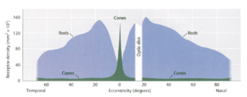
Distribution of Rods and Cones
Human's visual acuity varies across the visual field, with the region of highest acuity occurring at the center of gaze, called the fovea, and the lowest at the periphery. The fovea contains a higher concentration of cones than anywhere else on the retina and does not contain any rods. In order to achieve the highest possible acuity in the fovea, the cones are especially small and their retinal ganglion cells have small receptive fields. Peripheral ganglion cells have much larger receptive fields than foveal retinal ganglion cells. The distribution of rods and cones can be seen in the diagram below where the peak in cones corresponds to the fovea and the left and right edges of the plot correspond to peripheral vision:
The dramatic reduction in acuity associated with peripheral vision is not an evolutionary failure. Rather, it is the result of an architecture that serves a different function. The fovea is concerned with high spatial frequency information—fine details—while the periphery is used to evaluate low sptial frequrency information—coarse information, like blurry shadows along with the spatial scene taken as a whole.
When an artist creates a two-dimensional representation of a scene, the resolution across the surface may or may not match typical visual experience. If the resolution is homogenously conveyed in brutal detail, the image can loose its sense of vitality. The static image may be about a hyperactive moment (such as a battle scene), but without the appeal to our peripheral vision the image fails to achieve an accurate sense of liveliness.
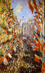
Impressionist painters like Monet overcome this problem by utilizing coarse information in a way that resonates well with the lived experience of a moment in time. See below:
|
|
Boulevard des Capucines, 1873
Claude Monet |
Rue Montorgueil in Paris, Festival of June 30, 1878, 1878
Claude Monet |
Leonardo DaVinci's Mona Lisa(credit) seems to live and breath . Take a look at her mouth. Now look elsewhere on the painting, at her eyes or the background, then return to her mouth. Do you notice anything peculiar about her smile as you scan your eyes back and forth? Margaret Livingstone has pointed out that her smile grows more apparent every time you shift your gaze away from it. That is, when viewed from your periphery, her smile is clear, but when viewed from the fovea it is not. Generally, facial expressions may be more obvious at lower spatial frequencies because they operate at the level of deep tissue. Research suggests that fine details are more important for identifying a specific person, whereas coarse information communicates the person's facial expression.